Computed Micro Tomography (Micro CT)
Micro-CT (Micro Computed Tomography)
High-Resolution Internal Structure Imaging and Analysis System
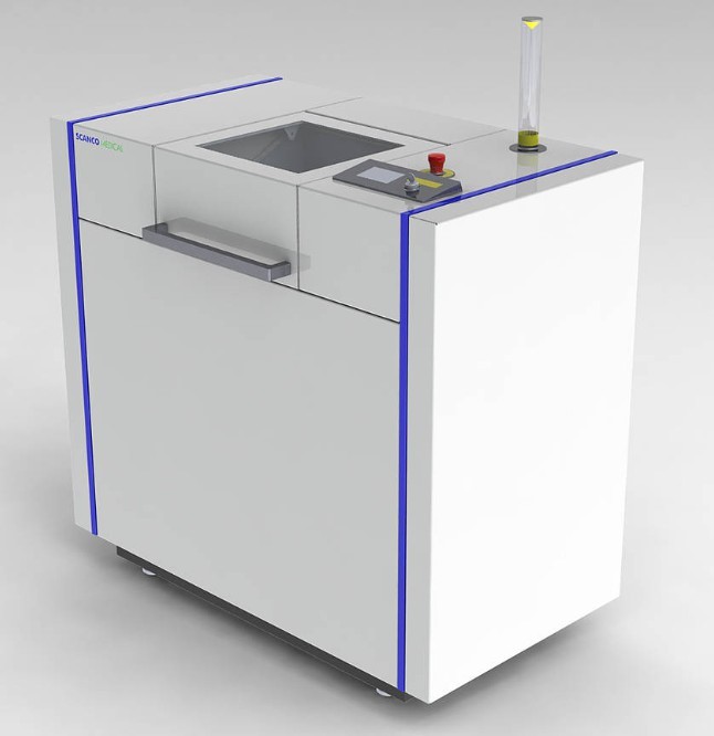
SCANCO MEDICAL µCT 50 device
Description
Micro-CT can create a three-dimensional (3D) model with images taken from the cross-sections of the material using X-rays without damaging the material. Analysis of the internal structure of materials such as composites, polymers, biological materials (bones, teeth, cartilage tissue, ...), bone morphometric analysis and micro-level imaging of up to 4 different materials in a sample can be performed. Apart from these features; by the Cooling/Heating Stage, the temperature can be decreased to 15°C below or increased to 15°C above the ambient temperature and analyzed under these conditions. The Compression/Tension Device is capable of analyzing the material under certain compression and tension conditions.
Key Features
- Ability to visualize up to 4 different material phases
- Non-destructive internal structure analysis
- Measurement under heating/cooling and loading/tension conditions
Application Areas
- Dentistry
- Biomedical
- Materials Science
- Geology
- Paleontology
- Food Industry
Device Configuration and Technical Specifications
Model: SCANCO MEDICAL µCT 50
Maximum Scanning Size: 48 x 100 mm (ØxL)
Maximum Sample Size: 50 x 120 mm (ØxL)
Resolution: 500 nm / <2 µm
Heating / Cooling Stage
- Maximum sample diameter: 8 mm
- Maximum height: 15 mm
- Maximum cooling capability: -15°C
- Maximum heating capability: +15°C
Compression / Tension Device
- Maximum Load/Tension: 450 N
- Maximum sample diameter: 23 mm
- Maximum height: 25 mm
- Maximum excursion: 8 mm
Please log in to the Erasis system to submit an analysis request.


